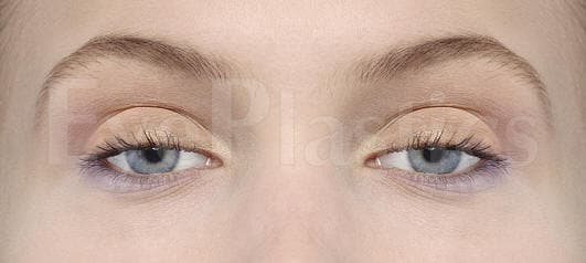
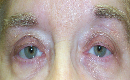
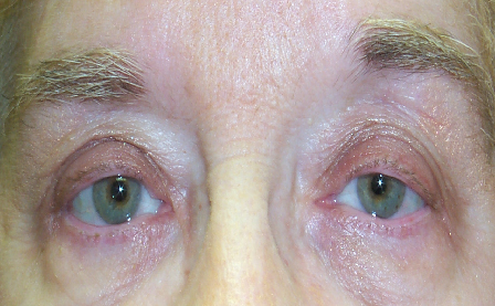
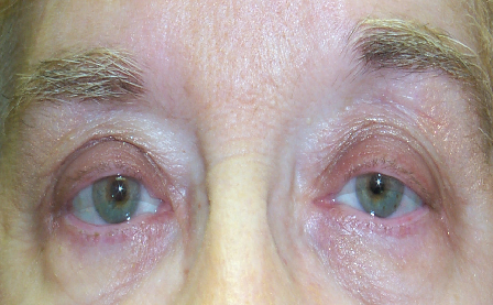
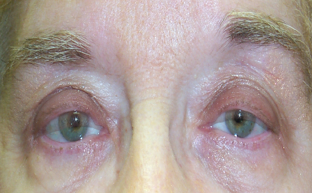
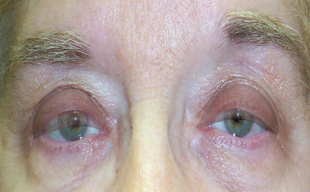
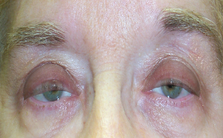
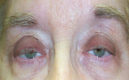
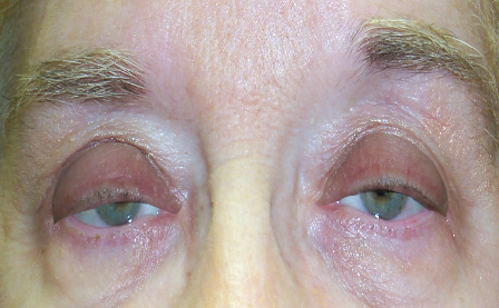
- Ptosis is also called blepharoptosis
- It differs from dermatochalasis (excess upper eyelid skin) — though both conditions frequently coexist
- A drooping lid that crosses the pupil reduces the superior visual field, causes eyebrow strain and headaches, and in children can lead to amblyopia (“lazy eye”)
For a detailed guide to levator anatomy, Müller’s muscle, and the tarsal plate, see our dedicated Anatomy Overview page.
Types & Causes of Ptosis
Ptosis is grouped by cause — age-related (aponeurotic), congenital, and neurologic. Recognizing the type guides both the work-up and the operation.
Explore Ptosis
Ptosis ranges from age-related drooping to conditions present from birth, each evaluated and treated differently. Explore each in depth:
Evaluation & DiagnosisMRD, levator function, the phenylephrine test, and Hering's law.
Treatment & SurgeryMüller's-muscle resection, levator advancement, frontalis sling, Upneeq.
Acquired PtosisThe common adult, age-related (aponeurotic) droopy eyelid and its causes.
Congenital PtosisA droopy lid from birth — and protecting a child's developing vision.
Horner's SyndromeMild ptosis with a small pupil and reduced facial sweating.
Marcus Gunn Jaw-WinkA congenital ptosis where the eyelid lifts as the jaw moves.
Ptosis & BlephLow lid margin (ptosis) vs. excess skin — and treating them together.
Symptoms of a Droopy Eyelid
Ptosis is more than a cosmetic concern — because the lid sits in the line of sight, it steadily narrows the field of view. Common signs include:
- A visibly lower eyelid on one or both sides, often described as looking tired or sleepy
- Difficulty reading or a sense that the upper part of your vision is cut off
- Chronically raised eyebrows and forehead ache from the frontalis muscle straining to lift the lids
- Tipping the head back or lifting the lid with a finger to see
- In children, a persistently drooping lid that can threaten visual development
Types & Causes in Depth
Identifying why the lid droops determines the correct operation, so surgeons classify ptosis by mechanism:
- Aponeurotic (age-related): The most common adult form. The levator tendon stretches or detaches from the tarsal plate with age, contact-lens wear, or after eye surgery. Levator strength is usually preserved. See Acquired Ptosis.
- Congenital: Present from birth, usually from a poorly developed levator muscle. Because a lid covering the pupil can cause amblyopia (“lazy eye”) in roughly 30% of cases, early evaluation is essential. See Congenital Ptosis.
- Neurogenic: A nerve-signal problem — third-nerve palsy, Horner’s syndrome (ptosis with a small pupil), or myasthenia gravis (fatigable, variable ptosis).
- Myogenic: The muscle itself is diseased, as in chronic progressive external ophthalmoplegia.
- Mechanical & synkinetic: A lid weighed down by mass or scarring, or the Marcus Gunn jaw-wink, where the lid lifts with jaw movement.
Ptosis (a low lid margin) is distinct from dermatochalasis (excess upper-lid skin), though the two frequently coexist — see Ptosis vs. Blepharoplasty.
How Ptosis Is Diagnosed
A focused eyelid exam measures a few key numbers that drive both the diagnosis and the surgical plan:
- Margin reflex distance (MRD-1): the gap from the central corneal light reflex to the upper-lid margin — normally about 4–5 mm. Ptosis is generally present when MRD-1 is 2 mm or less.
- Levator function: upper-lid travel from down- to up-gaze, graded good (≥10 mm), fair (5–9 mm), or poor (≤4 mm). This single measurement largely dictates which operation is chosen.
- Phenylephrine (Neo-Synephrine) test: drops that stimulate Müller’s muscle predict the response to a Müller’s-muscle conjunctival resection.
- Visual-field testing: taped vs. untaped fields document functional (insurance-qualifying) visual obstruction.
Full detail is on our Ptosis Evaluation page.
Treatment Options
Treatment is matched to the cause and to levator function. The main options:
- Levator advancement / resection: the workhorse repair when levator function is fair-to-good — the stretched muscle is reattached and tightened.
- Müller’s-muscle conjunctival resection (MMCR): an internal, no-external-scar option for mild ptosis with a positive phenylephrine test.
- Frontalis sling: for poor levator function (including many congenital cases), the lid is connected to the forehead muscle so the brow does the lifting.
- Oxymetazoline 0.1% (Upneeq) drops: a non-surgical daily eyedrop that lifts the lid a millimeter or two by stimulating Müller’s muscle — useful for mild ptosis or patients not ready for surgery.
See Ptosis Treatment & Surgery for how each is chosen and performed.
Cost & Insurance
Ptosis repair is often functional rather than cosmetic: when a formal visual-field test shows the lid obstructs your superior vision, the repair is frequently covered by insurance with prior authorization and photographs. If ptosis repair is combined with a cosmetic blepharoplasty, the skin (blepharoplasty) portion is billed separately as cosmetic. Ask the office for an itemized estimate and to verify coverage before surgery. The measurements insurers require, self-pay prices, and combined-billing rules are covered in Ptosis Surgery Cost & Insurance, part of our Eyelid Surgery Cost & Insurance guide.
Choosing an Oculoplastic Surgeon
Millimeters decide the result in ptosis surgery, and lid height must be balanced against the fellow eye (Hering’s law can unmask ptosis on the other side after repair). This is best handled by an oculoplastic surgeon with combined ophthalmology and eyelid-plastic training; ASOPRS fellowship-trained surgeons perform these repairs routinely.
Recovery & Results
Ptosis repair is typically an outpatient procedure under local anesthesia with light sedation. Bruising and swelling settle over 1–3 weeks, and because swelling shifts lid height, the final position is judged at a few weeks to a few months. Most repairs are durable for years; a minority need a minor height adjustment, which is a normal and expected part of fine-tuning millimeter-level symmetry.
Matching the Operation to Levator Function
The single measurement that drives the surgical plan is levator function — how far the upper lid travels from down-gaze to up-gaze. It sorts patients into three groups, each with a preferred repair:
- Good function (≥ 10 mm): the muscle works well and only needs tightening — a levator advancement, or an internal, no-external-scar Müller’s-muscle conjunctival resection (MMCR) when a phenylephrine test is positive.
- Fair function (5–9 mm): usually a levator advancement, with the amount of tightening titrated to the measured function.
- Poor function (≤ 4 mm): the muscle cannot lift the lid reliably, so a frontalis sling connects the lid to the forehead muscle and the brow does the lifting — the standard choice for most severe congenital cases.
Two measurements refine it further: MRD-1 (light-reflex to upper-lid margin, normally about 4–5 mm; ptosis at 2 mm or less) sets how much lift is needed, and Hering’s law warns that repairing one lid can unmask ptosis on the other — which an experienced surgeon accounts for before operating rather than discovering afterward. See the full work-up on our Ptosis Evaluation page and the operative detail on Ptosis Treatment.
Continue Reading — Complete Ptosis Guide
- Ptosis Evaluation & Diagnosis
- Ptosis Treatment & Surgery
- Acquired (Age-Related) Ptosis
- Congenital Ptosis
- Horner’s Syndrome
- Marcus Gunn Jaw-Wink
- Ptosis vs. Blepharoplasty